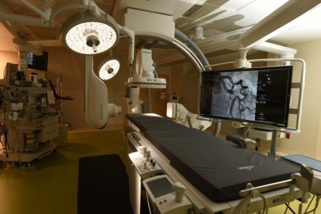
Nicklaus Children’s Hospital is the second pediatric facility in the world to use new medical technology in cardiac procedures that is similar to global positioning systems (GPS) that motorists use to determine their vehicles’ locations on a map.
The new equipment is part of the hospital’s new state-of-the-art cardiac catheterization lab, in which minimally invasive catheter-based procedures are performed to treat and evaluate children with congenital heart defects.
The MediGuide Technology allows physicians to see the precise location of specially designed delivery tools and catheters enabled with miniature sensors to navigate within the heart. Using a low-powered electromagnetic field, the system enables physicians to see in real-time, a three-dimensional (3-D) image of these tools on pre-recorded fluoroscopy (a rapid series of X-ray images). Automatic adjustments are made to the pre-recorded images to compensate for changing heart rhythms, breathing and patient movement. By allowing the physician to track devices on pre-recorded fluoroscopy, MediGuide technology can help avoid additional fluoroscopy throughout the procedure, which in turn significantly reduces radiation exposure.
“The longstanding commitment of The Heart Program at Nicklaus Children’s is to minimize the cumulative impact on the children in our care,” said Dr. John Rhodes, medical director of cardiology. “The MediGuide Technology supports us in this effort by reducing exposure to radiation during procedures and enhancing patient safety. It reflects our commitment to investing in the latest technology to ensure the best possible outcomes and safest practices for the children we serve.”
The MediGuide Technology may be used to aid a physician in navigating delivery tools and catheters in the cardiac anatomy during implantable device procedures, such as cardiac resynchronization therapy (CRT) cases, which helps restore a patient’s heart rhythm back to normal and protects against sudden cardiac death for CRT device patients. MediGuide Technology also can be used in ablation procedures where radiofrequency (RF) energy is delivered to correct an arrhythmia (or irregular heart beat).
Why reducing fluoroscopy is important
The current standard practice for viewing devices in the heart uses continuous, live fluoroscopy, which is a rapid series of X-ray images taken throughout the course of the procedure. Worldwide, physicians perform several billion of these radiation-based imaging procedures annually and approximately one-third occur in cardiovascular patients.
While the benefits of imaging provided by fluoroscopy are important, studies show that the ionizing radiation from repeated exposure to fluoroscopy over time may compromise a person’s immune system, elevate the risk of developing cancer, or present other health hazards.
For more information about Nicklaus Children’s Hospital, visit www.nicklauschildrens.org.


